
Ventral Carotid Canal Dehiscence in a Kenyan Population: An Osteological Study
Mots-clés :
Carotid canal, Dehiscence, Petrous Internal Carotid ArteryRésumé
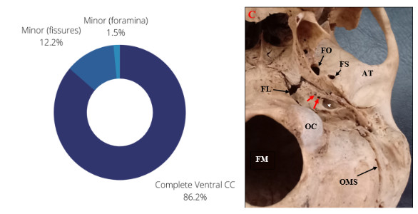
Background: The ventral surface of the carotid canal has previously been reported to be incomplete. Knowledge of this is important in the prevention of iatrogenic injuries during skull base procedures involving the petrous internal carotid artery and in the repair of carotid canal stenosis and fractures. Data on the prevalence of ventral carotid canal dehiscence locally remains scarce. Methods: 98 dry-sexed skulls were used to check for the presence of ventral carotid canal dehiscence. Data was analyzed using SPSS (Version 25.0) and frequency tables generated. Chi-square tests were used to assess associations with respect to side and sex. A p-value of ≤ 0.05 was considered significant. Results: Ventral (exocranial) dehiscence was present in 19.4% of skulls, with male skulls more likely to be dehiscent (p = 0.04). Minor dehiscence in the form of fissures was observed in 12.2% (24/196 sides), while holes were observed in 1.5% (3/196 sides). The rarer major dehiscence was not observed in this study. Bilateral dehiscence was observed in 8.2% (19 skulls). Conclusion: The ventral carotid canal was incomplete in 19.4% of Kenyan skulls. Procedures around the skull base should be done with caution to avoid inadvertent hemorrhage and vasospasm of the petrous internal carotid artery.
Références
Quint DJ, Silbergleit R, Young WC. Absence of the carotid canals at skull base CT. Radiology. 1992 Feb;182(2):477-81.
Hearst MJ, Kadar A, Keller JT, Choo DI, Pensak ML, Samy RN. Petrous carotid canal dehiscence: an anatomic and radiographic study. Otology & Neurotology. 2008 Oct 1;29(7):1001-4.
Pastor Vázquez JF, Gil Verona JA, García Porrero M. Carotid canal dehiscence in the human skull. Neuroradiology. 1999 Jun;41(6):447-9.
Toll EC, Browning M, Shukla R, Rainsbury JW. Cartilaginous Eustachian tube length and carotid canal dehiscence in children: a radiological study. European Archives of Oto-Rhino-Laryngology. 2018 Nov;275(11):2675-82.
Aoun MA, Nasr AY, Abdel Aziz AM. Morphometric Study of the Carotid Canal. Life Sci J 2013; 10(3):2559-2562.
Calgüner E, Turgut HB, Gözil R, Tunç E, Sevim A, Keskil S. Measurements of the carotid canal in skulls from Anatolia. Cells Tissues Organs. 1997;158(2):130-2.
Vidya CS, Shamasundar NM. Study of morphometry of carotid canal in skulls of South Indian origin. Journal of Clinical and Diagnostic Research: JCDR. 2015 Feb;9(2):AC16.
Osborn RE, Mojtahedi S, Hay TC, Dewitt JD. Internal carotid artery hypoplasia. Computerized radiology. 1986 Nov 1;10(6):283-7.
Inamasu J, Guiot BH. Iatrogenic carotid artery injury in neurosurgery. Neurosurgical review. 2005 Oct;28(4):239-47.
AlQahtani A, Castelnuovo P, Nicolai P, Prevedello DM, Locatelli D, Carrau RL. Injury of the internal carotid artery during endoscopic skull base surgery: prevention and management protocol. Otolaryngologic Clinics of North America. 2016 Feb 1;49(1):237-52
Téléchargements
Publiée
Comment citer
Numéro
Rubrique
Licence
(c) Tous droits réservés East African Journal of Neurological Sciences 2023

Ce travail est disponible sous licence Creative Commons Attribution - Pas d'Utilisation Commerciale - Pas de Modification 4.0 International.
