
Prevalence of the Posterior Ponticulus and Arcuate Foramen in a Kenyan Population: A Cross-Sectional Radiological Study
Keywords:
Arcuate Foramen, Posterior Ponticulus, Craniovertebral JunctionAbstract
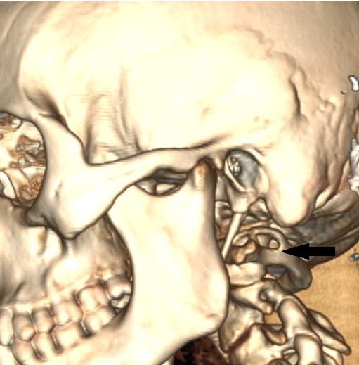
Background: Complete posterior ponticuli and the resultant arcuate foramen may compress the third part of the vertebral artery and the sub occipital nerve as they cross the neural arch of the atlas. This may manifest with neurological deficits and also complicate surgical access to lesions in the craniovertebral junction. The prevalence of the ponticulus has been shown to have sexual dimorphism and population differences. The aim of this study was to determine its prevalence and distribution across the sexes in the study population. Methodology: This was a cross-sectional Survey of 116 patients (equal numbers of males and females) done between August and October 2021 at the Radiology Department of Kenyatta National Hospital after ethical approval. Far lateral sagittal Computed tomography scans of the Craniovertebral junction and 3D reconstructions were analyzed in PACS software. The presence and completeness of the posterior ponticulus and arcuate foramen was determined. Chi square test was used to analyze gender differences in their distribution. Results: The prevalence of complete posterior ponticuli was at 15.5%, with unilateral ponticuli more common than bilateral ones (twelve versus six). There were no significant gender differences in terms of distribution. Conclusion: Variations of the posterior arch of the atlas are relatively high in the studied population, necessitating a low threshold for imaging before surgery, and high index of suspicion in neurological deficits involving the posterior circulation of the brain
Downloads
References
Sanchis-Gimeno JA, Blanco-Perez E, Perez-Bermejo M, Llido S, Nalla S. Retrotransverse foramen of the atlas: prevalence and bony variations. European Spine Journal. 2018 Jun;27(6):1272–7.
Cossu G, Terrier LM, Destrieux C, Velut S, François P, Zemmoura I, et al. Arcuate foramen: “Anatomical variation shape or adaptation legacy?” Surgical and Radiologic Anatomy. 2019 May;41(5):583–8.
Romanus t, tovi a. A variation of the atlas. Roentgenologic incidence of a bridge over the groove of the atlas for the vertebral artery. Acta Radiol Diagn (Stockh). 1964 Jul;2:289-97. doi: 10.1177/028418516400200403. PMID: 14198649.
Hiroli W, Gadade V. Radiological study of foramen arcuale: implications for screw insertion via posterior arch for fixation of C1 vertebrae in atlantoaxial instability using plain radiograph. Perspectives in Medical Research. 2023 Aug;11(2):61–6.
Dubey A, Rustagi SM, Prakash S, Dhuria R. Arcuate Foramen: An Anatomic Variant of Atlas Vertebra and Its Clinical Considerations. Journal of Medical Academics. 2023 Jun;6(1):3–7.
Sanchis-Gimeno JA, Ercan I, Llido S, Toluk Ö, Çini NT, Ozdemir ST, et al. Arcuate foramen prevalence in South African subjects: A cadaveric study based on 120 atlas vertebrae. Translational Research in Anatomy. 2023 Nov;33.
Karau PB, Ogengo JA, Hassanali J, Odula P. Anatomy and prevalence of atlas vertebrae bridges in a kenyan population: An osteological study. Clinical Anatomy. 2010;23(6):649–53.
P A, Henry BM, Jakub RP, Hsieh WC, Vikse J, Sanna B, et al. Prevalence of foramen arcuale and its clinical significance: a meta-analysis of 55,985 subjects. 2017;1–15.
Lamberty BG, Zivanović S. The retro-articular vertebral artery ring of the atlas and its significance. Acta Anat (Basel). 1973;85(1):113-22. doi: 10.1159/000143987. PMID: 4197316.
Cirpan S, Yonguc GN, Edizer M, Mas NG, Magden AO. Foramen arcuale: a rare morphological variation located in atlas vertebrae. Surgical and Radiologic Anatomy. 2017 Aug;39(8):877–84.
Afsharpour S, Hoiriis KT, Fox RB, Demons S. An anatomical study of arcuate foramen and its clinical implications: A case report. Chiropractic and Manual Therapies. 2016;24(1):1–7.
Allen W. The Varieties of the Atlas in the Human Subject, and the Homologies of its Transverse Processes. J Anat Physiol. 1879 Oct;14(Pt 1):18-27. PMID: 17231305; PMCID: PMC1309913.
Wight S, Osborne N, Breen AC. Incidence of Ponticulus Posterior of the Atlas in Migraine and Cervicogenic Headache. 1999;22(1):13–5.
Taitz C, Nathan H. Some observations on the posterior and lateral bridge of the atlas. Acta Anat (Basel). 1986;127(3):212-7. doi: 10.1159/000146284. PMID: 3788469.
Palancar CA, García-Martínez D, Radovčić D, Llidó S, Mata-Escolano F, Bastir M, et al. Krapina atlases suggest a high prevalence of anatomical variations in the first cervical vertebra of Neanderthals. Journal of Anatomy. 2020 Sep;237(3):579–86.
Murillo-llorente M, Perez-bermejo M, Nalla S. The Decreasing Prevalence of the Arcuate Foramen. World Neurosurgery. 2018;110:521–5.
Sanchís-Gimeno JA, Llidó S, Nalla S, Palancar CA. Arcuate Foramen in Chalcolithic and present subjects. Available from: https://www.researchgate.net/publication/332319013
Elliott RE, Tanweer O. The prevalence of the ponticulus posticus (arcuate foramen) and its importance in the Goel-Harms procedure: meta-analysis and review of the literature. World Neurosurg. 2014 Aug;82(1–2):e335-343.
Arslan D, Ozer MA, Govsa F, Kıtıs O. The Ponticulus Posticus as Risk Factor for Screw Insertion into the First Cervical Lateral Mass. World Neurosurgery. 2018 May;113:e579–85.
Wang W hao, Liu Z yong, Guo H cheng, Wang H. Multiple Fractures of Cervical Vertebrae Combined with Arcuate Foramen and Vertebral Artery Occlusion: A Case Report and Literature Review. Orthopaedic Surgery. 2021 Feb;13(1):360–5.
Hyun G, Allam E, Sander P, Hasiak C. The prevalence of congenital C1 arch anomalies. European Spine Journal. 2017;
Chen C hui, Chen Y kwan, Wang C kuo. Prevalence of ponticuli posticus among patients referred for dental examinations by cone-beam CT. The Spine Journal. 2015;15(6):1270–6.
Tubbs RS, Johnson PC, Shoja MM, Loukas M, Oakes WJ. Foramen arcuale: anatomical study and review of the literature. J Neurosurg Spine. 2007 Jan;6(1):31-4. doi: 10.3171/ spi.2007.6.1.6. PMID: 17233288.
Goel A, Laheri V. Plate and screw fixation for atlanto-axial subluxation. Acta Neurochir (Wien). 1994;129(1–2): 47–53.
Cho YJ. Radiological analysis of ponticulus posticus in Koreans. Yonsei Medical Journal. 2009;50(1):45–9.
Van P, Lacy PD, Konopková R, Lacman J, Bene V. Vertebral artery and osseous anomalies characteristic at the craniocervical junction diagnosed by CT and 3D CT angiography in normal Czech population : analysis of 511 consecutive patients. 2016;
Sharma V, Chaudhary D, Mitra R. Prevalence of ponticulus posticus in Indian orthodontic patients. Dentomaxillofacial Radiology. 2010;39(5):277–83.
Rahman MA. Incidence of the Arcuate Foramen of Atlas Vertebra and its Morphometric Differences with the Ipsilateral Transverse Foramen in the Egyptian Population [Internet]. Vol. 90, Cairo Univ. 1397. Available from: www.medicaljournalofcairouniversity.net
Downloads
Published
License
Copyright (c) 2025 East African Journal of Neurological Sciences

This work is licensed under a Creative Commons Attribution-NonCommercial-NoDerivatives 4.0 International License.
