
A Radiological Assessment of the Morphology and Morphometry of the Vidian Canal in a select Kenyan Population
Keywords:
vidian canal, medial pterygoid plate, endoscopic endonasal approachesAbstract
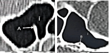
Background: The increased use of endoscopic approaches to the skull base has necessitated the identification of landmarks that assist in avoiding iatrogenic injury to structures within the skull base. The vidian canal (VC) is an established landmark for the petrous portion of the internal carotid artery (ICA). Although population differences have been reported in the literature, the area remains relatively unexplored in African populations. Materials and methods: Axial and coronal sections of 96 high-resolution computed tomography scans of Kenyan skulls were analyzed to determine VC morphometry. The VC length was determined using axial sections, and the VC - medial pterygoid plate (MPP) distance was determined using coronal sections. Results: The mean VC length studied was 16.50±2.30mm (10.50-22.40mm). No statistically significant differences were noted between sides (p=0.686) or sexes (p= 0.826, 0.593). The mean VC-MPP distance was 9.60±2.70 mm (2.40-21.40mm). No statistically significant differences were noted between sides (p=0.237) or sexes (p=0.886,0.850). The relational configurations of the VC to the SS were noted as follows: Type I (wholly within the cavity of the SS)-21.35%, Type II (VC on the floor of the SS or partially protruding into the SS) -76.57%, Type III (VC within the sphenoid corpus)- 2.08%. No significant correlation was noted between the type of VC configuration described and the side of the skull studied (p= 0.499). However, a significant correlation was noted between the type of VC configuration and sex (p=0.001). Conclusion: The increased prevalence of type I VCs among males indicates an increased risk during transsphenoidal surgical approaches in them.
Downloads
References
Kurt H, Bozkurt P, Bilecenoglu B, Kolsuz M, Orhan K. Morphometric analysis of vidian canal and its relations with surrounding anatomic structures by using cone beam computed tomography. Folia morphologica. 2019 Sep 1;
Açar G, Çiçekcibaşı AE, Çukurova İ, Özen KE, Şeker M, Güler İ. The anatomic analysis of the vidian canal and the surrounding structures concerning vidian neurectomy using computed tomography scans. Braz J Otorhinolaryngol. 2019 Apr;85(2):136–43.
Bahşi İ, Orhan M, Kervancıoğlu P, Yalçın ED. The anatomical and radiological evaluation of the Vidian canal on cone-beam computed tomography images. Eur Arch Otorhinolaryngol. 2019 May;276(5):1373–83.
Benner D, Hendricks BK, Benet A, Lawton MT. Eponyms in Vascular Neurosurgery: Comprehensive Review of 11 Arteries. World Neurosurgery. 2021 Jul 1;151:249–57.
Kassam AB, Vescan AD, Carrau RL, Prevedello DM, Gardner P, Mintz AH, et al. Expanded endonasal approach: vidian canal as a landmark to the petrous internal carotid artery. J Neurosurg. 2008 Jan;108(1):177–83.
Mato D, Hirono S, Yokota H, Martino J, Saeki N. The Vidian Canal: Radiological Features in Japanese Population and Clinical Implications. Neurologia medico-chirurgica. 2015 Jan 15;55:71–6.
Kasemsiri P, Solares CA, Carrau RL, Prosser JD, Prevedello DM, Otto BA, et al. Endoscopic endonasal transpterygoid approaches: anatomical landmarks for planning the surgical corridor. Laryngoscope. 2013 Apr;123(4):811–5.
Yazar F, Cankal F, Haholu A, Kilic C, Tekdemir I. CT evaluation of the Vidian canal localization. Clinical anatomy (New York, NY). 2007 Oct 1;20:751–4.
Lee JC, Kao CH, Hsu CH, Lin YS. Endoscopic transsphenoidal vidian neurectomy. Eur Arch Otorhinolaryngol. 2011 Jun;268(6):851–6.
Martínez-Abadías N, Esparza M, Sjøvold T, González-José R, Santos M, Hernández M. Heritability of human cranial dimensions: comparing the evolvability of different cranial regions. J Anat. 2009 Jan;214(1):19–35.
Vescan AD, Snyderman CH, Carrau RL, Mintz A, Gardner P, Branstetter B, et al. Vidian canal: analysis and relationship to the internal carotid artery. Laryngoscope. 2007 Aug;117(8):1338–42.
Ucerler H, Aktan Ikiz ZA, Yoruk MD, Boduc E, Ozturk L. Morphometric assessment of important landmarks on skull intended for Vidian nerve surgery. Surg Radiol Anat. 2020 Sep;42(9):987–93.
Cheng Y, Gao H, Song G, Li Y, Zhao G. Anatomical study of pterygoid canal (PC) and palatovaginal canal (PVC) in endoscopic trans-sphenoidal approach. Surgical and Radiologic Anatomy. 2016 Jul 1;38.
Bidarkotimath S, S V, udyavar ajay. Vidian canal: Radiological anatomy and functional correlations. journal of morphological sciences. 2012 Jan 1;29:27–31.
Lakshman N, Viveka S, Thondupadath Assanar FB. Anatomical relationship of pterygoid process pneumatization and vidian canal. Brazilian Journal of Otorhinolaryngology [Internet]. 2020 Jul 21 [cited 2021 May 31]; Available from: https://www.sciencedirect.com/science/article/pii/S180886942030104X
Omami G, Hewaidi G, Mathew R. The neglected anatomical and clinical aspects of pterygoid canal: CT scan study. Surgical and radiologic anatomy : SRA. 2011 Mar 1;33:697–702.
Mohebbi A, Rajaeih S, Safdarian M, Omidian P. The sphenoid sinus, foramen rotundum and vidian canal: a radiological study of anatomical relationships. Braz J Otorhinolaryngol. 2017 Aug;83(4):381–7.
Yeğin Y, Çelik M, Altıntaş A, Şimşek BM, Olgun B, Kayhan FT. Vidian Canal Types and Dehiscence of the Bony Roof of the Canal: An Anatomical Study. Turk Arch Otorhinolaryngol. 2017 Mar;55(1):22–6.
Liu SC, Wang HW, Su WF. Endoscopic Vidian Neurectomy: The Value of Preoperative Computed Tomographic Guidance. Arch Otolaryngol Head Neck Surg. 2010 Jun 21;136(6):595.
Downloads
Published
License
Copyright (c) 2024 East African Journal of Neurological Sciences

This work is licensed under a Creative Commons Attribution-NonCommercial-NoDerivatives 4.0 International License.
