
An MRI-Based Normative Morphometric Atlas of the Corpus Callosum and Ventricular System in a Northwestern Nigerian Population: A Descriptive Cross-sectional Study
Schlagwörter:
Corpus callosum, Ventricular system, Brain morphometry, Normative data, NeuroanatomyAbstract
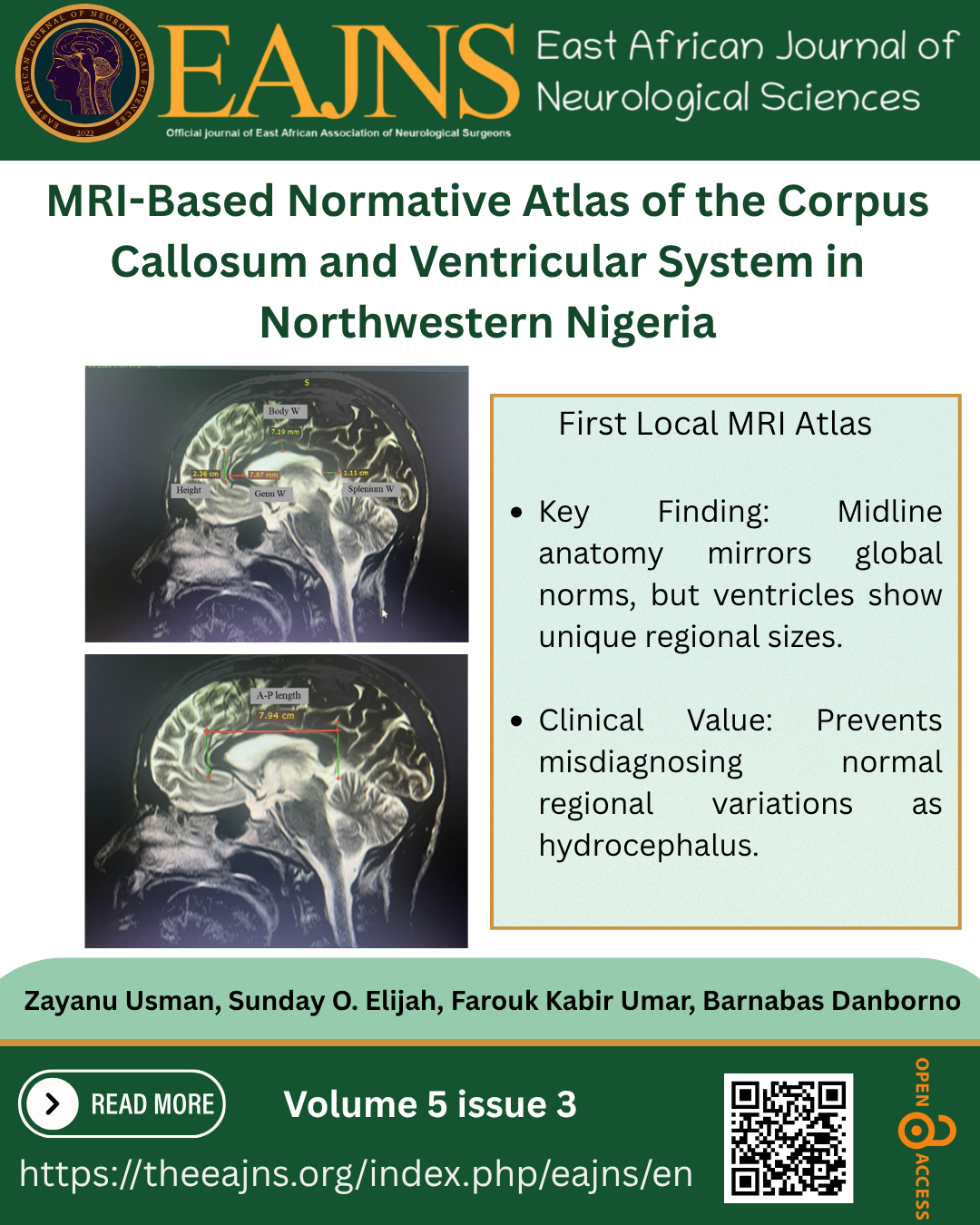
Background: Accurate morphometric definition of the corpus callosum (CC) and ventricular system is important for clinical neuroradiology and neuroanatomic research. Northwestern Nigeria lacks population-specific normative MRI data for adults, limiting accurate anatomical interpretation and diagnostic accuracy. Objective: To determine the MRI-based morphometric dimensions of the corpus callosum and ventricular system among neurologically normal adults in Northwestern Nigeria. Methods: A retrospective cross-sectional study of 422 MRI scans of adults (18 to 80 years) from Usmanu Danfodiyo University Teaching Hospital, Sokoto. Using sagittal T1-weighted and axial T2-weighted sequences, CC (rostrum, genu, body, and splenium) and ventricular subregions (lateral, third, and fourth ventricles; frontal and posterior horns) were identified. Measurements followed standardised protocols. Sex-stratified descriptive statistics and percentile-based reference ranges (2.5th-97.5th) were assessed. Results: The anteroposterior length of the CC was approximately 68.1 mm to 81.3 mm, and other sub-regions of the callosal region exhibited low dispersion, implying that there is a stable midline morphology. An increased range of variance was observed in the ventricular dimensions, with a mean lateral ventricular width of 8.01 ± 0.799 mm, a third ventricular width of 4.19 ± 0.869 mm, and a frontal horn width of 33.32 ± 0.264 mm. A normative percentile range of all the CC and ventricular measurements was established. Conclusion: This study presents the first comprehensive MRI-based normative morphometric atlas of the corpus callosum and ventricular system for adults in northwestern Nigeria. Such reference data promote anatomical understanding and aid accurate neuroradiological analysis in clinical practice.
Downloads
Literaturhinweise
1. Prendergast DM, Ardekani B, Ikuta T, John M, Peters B, DeRosse P, Wellington R, Malhotra AK, Szeszko PR. Age and sex effects on corpus callosum morphology across the lifespan. Human brain mapping. 2015 Jul;36(7):2691-702.
2. Lavrador JP, Ferreira V, Lourenço M, Alexandre I, Rocha M, Oliveira E, Kailaya-Vasan A, Neto L. White-matter commissures: a clinically focused anatomical review. Surgical and Radiologic Anatomy. 2019 Jun 1;41(6):613-24.
3. Ughwanogho UO, Taber KH, Chiou-Tan FY. Special anatomy series: Updates in structural, functional, and clinical relevance of the corpus callosum: What new imaging techniques have revealed. Journal of the International Society of Physical and Rehabilitation Medicine. 2022 Jul 1;5(3):81-9.
4. Ruigrok AN, Salimi-Khorshidi G, Lai MC, Baron-Cohen S, Lombardo MV, Tait RJ, Suckling J. A meta-analysis of sex differences in human brain structure. Neuroscience & Biobehavioral Reviews. 2014 Feb 1;39:34-50.
5. Toga AW, Thompson PM. Mapping brain asymmetry. Nature Reviews Neuroscience. 2003 Jan;4(1):37-48.
6. Patra A, Kaur H, Chaudhary P, Ravi KS, Asghar A, Walocha J, Iskra T, Blaszczyk M. Quantitative Analysis of Cerebral Ventricular Dimensions in the Adult Population of Northern India: A Cross-Sectional Study Using Magnetic Resonance Imaging: Quantitative Analysis of Cerebral Ventricular Dimensions. La Clinica Terapeutica. 2025 Feb 17;176(1).
7. Evans WA. An encephalographic ratio for estimating ventricular enlargement and cerebral atrophy. Archives of Neurology & Psychiatry. 1942 Jun 1;47(6):931-7.
8. Nestor SM, Rupsingh R, Borrie M, Smith M, Accomazzi V, Wells JL, Fogarty J, Bartha R, Alzheimer's Disease Neuroimaging Initiative. Ventricular enlargement as a possible measure of Alzheimer's disease progression validated using the Alzheimer's disease neuroimaging initiative database. Brain. 2008 Sep 1;131(9):2443-54.
9. Takeda S, Hirashima Y, Ikeda H, Yamamoto H, Sugino M, Endo S. Determination of indices of the corpus callosum associated with normal aging in Japanese individuals. Neuroradiology. 2003 Aug;45(8):513-8.
10. Sun L, Qin W, Liang X, Wang C, Men W, Duan Y, Fan XR, Cai Q, Qiu S, Wang M, Gong Q. Population-specific brain charts reveal Chinese-Western differences in neurodevelopmental trajectories. bioRxiv. 2025 Jun 18. https://doi.org/10.1101/2025.06.17.659820
11. Junle Y, Youmin G, Yanjun G, Mingyue M, Qiujuan Z, Min X. A MRI quantitative study of corpus callosum in normal adults. Journal of Medical Colleges of PLA. 2008 Dec 1;23(6):346-51.
12. Bakken TE, Dale AM, Schork NJ, Alzheimer’s Disease Neuroimaging Initiative. A geographic cline of skull and brain morphology among individuals of European Ancestry. Human heredity. 2011 Aug 17;72(1):35-44.
13. Bonfili N, Barbeito‐Andrés J, Bernal V, Hallgrímsson B, Gonzalez PN. Morphological correspondence between brain and endocranial surfaces in mice exposed to undernutrition during development. Journal of Anatomy. 2022 Jul;241(1):1-2. doi:10.1111/joa.13639.
14. Prasad S. Variability in neuroimaging research is not always wrong. Nature Human Behaviour. 2023 Dec;7(12):2048-9. https://doi.org/10.1038/s41562-023-01767-7
15. Ajare EC, Campbell FC, Mgbe EK, Efekemo AO, Onuh AC, Nnamani AO, Okwunodulu O, Ohaegbulam SC. MRI-based morphometric analysis of corpus callosum dimensions of adults in Southeast Nigeria. Libyan Journal of Medicine. 2023 Dec 31;18(1):2188649. https://doi.org/10.1080/19932820.2023.2188649
16. Allouh MZ, Al Barbarawi MM, Ali HA, Mustafa AG, Alomari SO. Morphometric analysis of the corpus callosum according to age and sex in Middle Eastern Arabs: racial comparisons and clinical correlations to autism spectrum disorder. Frontiers in Systems Neuroscience. 2020 Jun 23;14:30. https://doi.org/10.3389/fnsys.2020.00030
17. Ominde B, Enaohwo M, Ikubor J, Denise O, Jeremiah O, Omoro O, Igbigbi P. MRI-based morphometric study of the corpus callosum in Nigerian adults. Niger Health Journal 2025;25(1):357–364 . https://doi.org/10.71637/tnhj.v25i1.989
18. David LK, Vidona WB. Anatomical evaluation of the craniometric points and dimensions among adult’s populations of the South-Eastern Nigerians and its implication for intracranial surgical procedures. Glob J Medical Clin Case Rep. 2021;8(3):116-9. https://dx.doi.org/10.17352/2455-5282.000143
19. Evans AC, Janke AL, Collins DL, Baillet S. Brain templates and atlases. Neuroimage. 2012 Aug 15;62(2):911-22. https://doi.org/10.1016/j.neuroimage.2012.01.024
20. Mazziotta J, Toga A, Evans A, Fox P, Lancaster J, Zilles K, Woods R, Paus T, Simpson G, Pike B, Holmes C. A probabilistic atlas and reference system for the human brain: International Consortium for Brain Mapping (ICBM). Philosophical Transactions of the Royal Society of London. Series B: Biological Sciences. 2001 Aug 29;356(1412):1293-322. https://doi.org/10.1098/rstb.2001.0915
21. Zilles K, Amunts K. Individual variability is not noise. Trends in cognitive sciences. 2013 Apr 1;17(4):153-5. https://doi.org/10.1016/j.tics.2013.02.003
22. Tang Y, Hojatkashani C, Dinov ID, Sun B, Fan L, Lin X, Qi H, Hua X, Liu S, Toga AW. The construction of a Chinese MRI brain atlas: a morphometric comparison study between Chinese and Caucasian cohorts. Neuroimage. 2010 May 15;51(1):33-41. https://doi.org/10.1016/j.neuroimage.2010.01.111
23. Fischl B. FreeSurfer. Neuroimage. 2012 Aug 15;62(2):774-81. https://doi.org/10.1016/j.neuroimage.2012.01.021
24. Thompson PM, Stein JL, Medland SE, Hibar DP, Vasquez AA, Renteria ME, Toro R, Jahanshad N, Schumann G, Franke B, Wright MJ. The ENIGMA Consortium: large-scale collaborative analyses of neuroimaging and genetic data. Brain imaging and behavior. 2014 Jun;8(2):153-82. https://doi.org/10.1007/s11682-013-9269-5
25. Krishna VB, Sekhar KC, Pravallika D, Chandana TH. Corpus callosal morphometry in mid-sagittal plane MRI in patients of different age groups: a retrospective study. Int J Anat Radiol Surg. 2022;11(2):31-3. https://www.doi.org/10.7860/JCDR/2022/51289/2780.
26. Cesana, B. M., Antonelli, P., & Ferraro, S. (2025). Critical appraisal of the CLSI guideline EP09c “measurement procedure comparison and bias estimation using patient samples”. Clinical Chemistry and Laboratory Medicine (CCLM), 63(3), 507-514.
27. Charan J, Biswas T. (2013). How to calculate sample size for different study designs in medical research. Indian Journal of Psychological Medicine, 35(2), 121–126
Downloads
Veröffentlicht
Erklärung zur Datenverfügbarkeit
We are yet to make our data available to the general public. this may later
Lizenz
Copyright (c) 2026 East African Journal of Neurological Sciences

Dieses Werk steht unter der Lizenz Creative Commons Namensnennung - Nicht-kommerziell - Keine Bearbeitungen 4.0 International.
